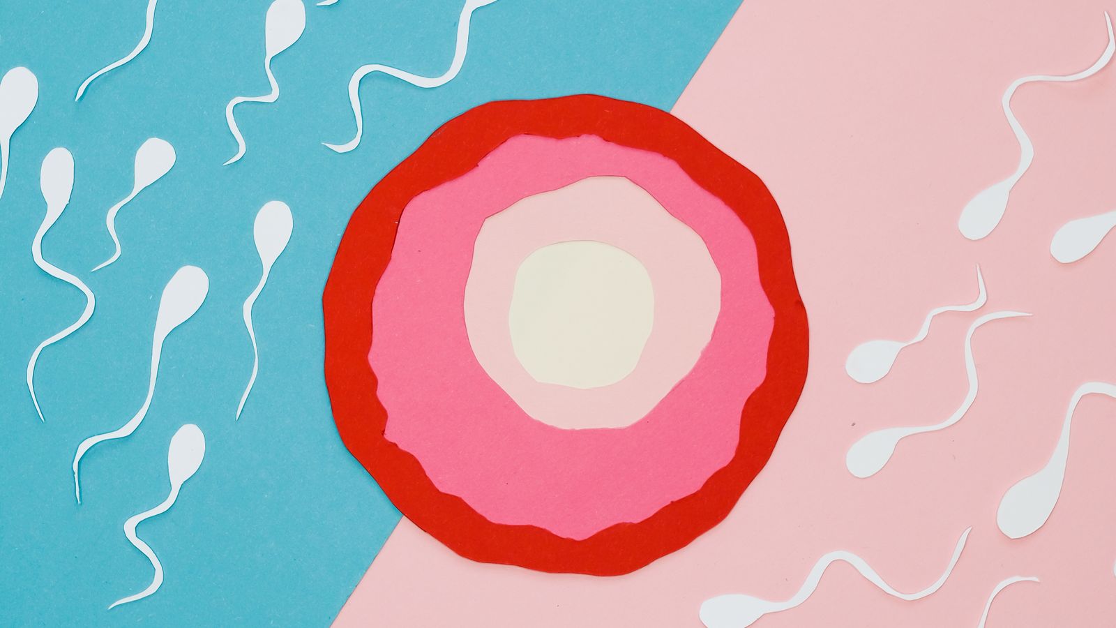
Quick answer
A mature human egg (ovum) is approximately 0.1 millimeter across — about 100 micrometers. That makes it the largest cell in the human body and, uniquely among human cells, just visible to the naked eye. For comparison, a sperm cell is about half a millimeter long total but its head — which carries the DNA — is only 5 micrometers, roughly 20 times smaller than the egg in diameter and thousands of times smaller by volume.
The human egg — the ovum — is one of the strangest cells in the body. It's the largest single cell you have, it sits at a precise size that puts it right at the edge of visibility, and every one you'll ever release was already inside you when you were still in the womb. If you've ever wondered how big a human egg actually is, or how it compares to sperm, or where the "400 eggs in a lifetime" number comes from, this is the honest breakdown.
We'll walk through the actual dimensions, useful comparisons (grain of sand, period at the end of a sentence, hair diameter), the anatomy of the egg, how eggs develop from primordial follicles to the mature Graafian follicle that ovulates, egg count from birth through menopause, and what happens to the egg at ovulation. Along the way we'll dispel a few common misconceptions.
A quick source note: we're the team behind Vyve, a private AI cycle tracker with automatic BBT/HRV integration from wearables. This piece is educational, not medical advice, and none of it is a diagnostic tool. If you're navigating fertility questions personally, please loop in a clinician.
On this page
- The actual size of a human egg
- Compared to everyday things
- Egg vs sperm: the scale mismatch
- Anatomy of a mature egg
- From primordial follicle to Graafian
- How many eggs across a lifetime
- What happens at ovulation
- Egg quality vs egg quantity
- Common myths about eggs
- When to see a doctor
- How Vyve helps you track ovulation
- Frequently asked questions
The actual size of a human egg
A mature, ready-to-ovulate human egg measures approximately 100 micrometers in diameter. In more familiar units that's 0.1 millimeter, or about a tenth the width of a typical sesame seed. This size is remarkably consistent — mature ova across healthy human ovaries all cluster tightly around 100 μm, with the entire cell (including its protective coats) topping out around 130-150 μm.
Two things make this number worth pausing on. First, it's tiny by everyday standards. If you imagine the period at the end of this sentence — the standard printed dot — that's roughly the same size, maybe a touch larger. Second, in the context of your body, this is enormous. Most human cells are 10 to 30 micrometers across. Red blood cells are about 7 μm. Even most nerve cell bodies (setting aside their long axons) are under 40 μm. A mature egg is roughly ten times the diameter of an average body cell, and by volume that's a thousandfold difference. It really is the biggest cell you have.
Why so big? Because the egg has to do work no other cell does. After fertilization, it supplies all the cellular machinery — mitochondria, ribosomes, membranes, mRNA, nutrients — for the first several days of embryonic development. Sperm bring DNA and essentially nothing else. Everything the early embryo does, it does using resources the egg already stockpiled during its long development in the ovary. That's what makes it so large.
Key takeaway
A mature human egg is ~100 micrometers (0.1 mm) across — the largest cell in the human body and just barely visible to the naked eye. It's big because it has to carry the cellular machinery for the entire early embryo.
Compared to everyday things
Numbers alone are hard to feel. Here's the egg's size sitting next to things you can picture.
| Object | Size | Relative to egg |
|---|---|---|
| Mature human egg | ~100 μm (0.1 mm) | Reference |
| Period at end of sentence (printed) | ~150-300 μm | Slightly larger |
| Fine human hair (diameter) | ~50-100 μm | About the same |
| Fine beach sand grain | ~100-500 μm | Similar to larger |
| Red blood cell | ~7 μm | ~14× smaller |
| Sperm cell (head only) | ~5 μm | ~20× smaller |
| Bacterium (E. coli) | ~2 μm | ~50× smaller |
The most useful mental image is probably the printed period, because you can look at one right now. A ripe human egg is about the same size as the dot at the end of this sentence when it's printed at typical body-text size. That's small — but it's not microscopic in the strict sense. You can, in the right light and knowing exactly where to look, actually see one.
Embryologists doing IVF confirm this in practice: they routinely spot individual eggs under low-magnification stereo microscopes without needing full high-power optics, and can even see them with the naked eye against the right background. That's essentially unheard-of for any other human cell.
Egg vs sperm: the scale mismatch
The egg-to-sperm comparison is dramatic, and it's one of the things people find most surprising about human reproduction. A sperm cell is about 50 micrometers long from head to tail tip. But almost all of that length is the tail, which is basically a whip for swimming. The sperm's head — the part that fuses with the egg and delivers the paternal DNA — is only about 5 μm across.
So the diameter comparison is roughly 100 μm (egg) to 5 μm (sperm head) — a 20-fold difference. But diameter understates things. Because both cells are roughly spherical or ovoid, the volume comparison is closer to 20 cubed, or around 8,000 times. A mature human egg has thousands of times the cellular volume of a sperm cell.
Sperm are essentially DNA delivery vehicles. The egg is where all the cellular substance of the early embryo comes from. That asymmetry is exactly why the egg is so enormous — and so precious.
This asymmetry drives a lot of the reproductive biology that follows. Because the egg is so large and metabolically expensive to produce, the ovary releases only one per cycle in most people (occasionally two — that's how fraternal twins happen). Because sperm are small and cheap, testes make them in the hundreds of millions and release tens to hundreds of millions per ejaculation. When we get to the topic of where fertilization happens, in our companion piece on where sperm fertilizes the egg, this asymmetry does a lot of the explanatory work.
Anatomy of a mature egg
A mature egg isn't just a single blob — it has distinct concentric layers, each with a job. From the inside out:
- The egg cell itself (oocyte). At the center is the actual cell, containing the nucleus with the maternal DNA and a large cytoplasm packed with organelles, nutrients, mRNA, and proteins the early embryo will need.
- Perivitelline space. A thin fluid-filled gap between the egg's plasma membrane and the next layer out. This is where the second polar body — a tiny leftover of egg cell division — sits.
- Zona pellucida. A thick, tough glycoprotein coat around the egg. Sperm must chemically and mechanically penetrate this layer to fertilize the egg. After fertilization the zona hardens (the "cortical reaction") to block additional sperm from entering.
- Corona radiata. A single tight layer of cumulus cells attached directly to the zona pellucida. These cells help support and guide the egg.
- Cumulus oophorus. A larger fluffy cloud of cumulus cells outside the corona radiata. These cells nourished the egg during its final maturation and help the fallopian tube's ciliated cells "grab" the egg after ovulation.
So when an egg is ovulated, it's really an egg complex: the egg cell itself is only about 100 μm, but the whole cumulus cloud makes the released package closer to 500 μm — visible to the naked eye without much difficulty. The whole complex is what gets swept from the ovary surface into the fallopian tube.
From primordial follicle to Graafian follicle
An egg doesn't start out at 100 μm. It grows — dramatically — over months, inside the structure that houses it in the ovary: the follicle. Understanding the follicle stages helps make sense of both the ovulation cycle and why fertility varies so much with age.
Primordial follicle. The starting state. A tiny immature egg surrounded by a single flat layer of granulosa cells. About 30 μm total. All of a baby girl's eggs are in this state at birth.
Primary and secondary follicles. The granulosa layer thickens, the egg grows, and additional supporting cells form. Now around 50-200 μm.
Antral (early tertiary) follicle. A fluid-filled cavity called the antrum forms inside the follicle. From this stage onward the follicle grows quickly. Several antral follicles are recruited at the start of each menstrual cycle — this is what's happening during the follicular phase.
Dominant follicle. Usually only one follicle "wins" the recruitment race in each cycle. It continues to grow while the others regress and are reabsorbed.
Graafian (preovulatory) follicle. The mature, ready-to-ovulate follicle. This is a fluid-filled sac up to 20 millimeters (2 cm) across containing the fully mature egg. That's 200 times the diameter of the egg itself. The follicle bulges out of the ovary surface — and when the LH surge arrives, it ruptures, releasing the egg and its cumulus complex.
The whole process from primordial follicle to ovulated egg takes many months — realistically about 6 to 8 months of gradual growth, with the final rapid phase happening during the current cycle. This is why lifestyle changes for fertility take time to show effects: the egg you'll ovulate three months from now was already well into its development phase months ago.
How many eggs across a lifetime
Here's a fact that surprises almost everyone: you were born with your entire lifetime supply of eggs, and the number has been decreasing ever since. Unlike sperm, which men produce continuously throughout life, eggs are not made after birth in humans. This is one of the most consequential facts in reproductive biology.
The numbers, in rough terms:
- Peak fetal life (~20 weeks gestation): approximately 6-7 million immature eggs — the highest number a person will ever have.
- Birth: approximately 1-2 million eggs. The huge drop from fetal peak is a normal, natural process called atresia in which most eggs simply degrade before birth.
- Puberty: approximately 300,000-400,000 eggs. Most eggs are lost to atresia between birth and puberty.
- Age 30: approximately 100,000-150,000 eggs remaining.
- Age 40: approximately 10,000-25,000 eggs remaining.
- Menopause: under 1,000 eggs — the functional threshold has been crossed and cycles stop.
Now the punchline: of those original million-plus, only about 400 eggs are ever ovulated across an entire lifetime. That's roughly one per cycle for about 30 years of reproductive life. Everything else — over 99.9% of the initial supply — is lost to atresia along the way. The body is continuously "shedding" eggs whether or not you're cycling, on the pill, pregnant, or otherwise. The pill doesn't preserve eggs; menopause happens on essentially the same timetable regardless.
Key takeaway
You're born with 1-2 million eggs and lose them continuously across life. Only about 400 are ever ovulated. This one-way math is why "fertility declines with age" is a biological fact, not a cultural narrative — and why timing matters if pregnancy is part of the plan.
What happens at ovulation
Ovulation itself is fast. In the hours after the LH surge peaks, enzymes weaken the wall of the Graafian follicle at a specific spot called the stigma. The follicle then ruptures — usually as a gentle release, sometimes with a small pop that a person can feel as mittelschmerz — and the mature egg plus its cumulus complex is pushed out onto the surface of the ovary.
Almost immediately, the fringe-like ends of the fallopian tube (the fimbriae) sweep over the ovary surface and catch the egg. Cilia on the inside of the tube then propel the egg toward the ampulla, which is the widest section of the fallopian tube and where fertilization actually happens. Our companion piece on where sperm fertilizes the egg walks through the rest of the journey in detail.
The egg's clock starts ticking immediately after release: it survives only about 12 to 24 hours before it can no longer be fertilized. If sperm are already waiting in the fallopian tube — which they can be, because they can survive up to five days in fertile mucus — fertilization can happen within minutes to hours. Otherwise the egg is lost, and the cycle proceeds to the luteal phase and eventually to the next period.
If you want to actually catch this in real time — noting when ovulation is happening in your own body — our guide to the signs of ovulation covers the mucus, temperature and LH surge signals that let you spot it.
Track ovulation honestly, on your device
Vyve reads BBT and HRV automatically from your Apple Watch, Whoop or Oura to identify ovulation and your fertile window — even on irregular cycles. Private, AI-powered, never sold.
Try Vyve todayEgg quality vs egg quantity
Egg size stays essentially constant across the reproductive years — a mature 25-year-old's egg and a mature 40-year-old's egg are the same 100 μm across. What changes with age is egg quality and egg quantity, and both matter for fertility.
Quantity declines steadily, as the numbers above show. Quality declines partly because the eggs remaining have been sitting in the ovary since before birth — the eggs still available at age 40 are the same eggs that were there in fetal life, forty years older. Older eggs are more likely to have chromosomal errors, which is the main biological reason miscarriage rates and chromosomal condition rates rise gradually with maternal age.
This is genuinely useful information, not a scare tactic. Fertility doesn't fall off a cliff at 35 — it declines gradually across the 30s and 40s — but the direction of travel is real, and knowing about it lets people plan. Egg freezing (technically oocyte cryopreservation) exists specifically because harvested eggs frozen at, say, 32 stay biologically 32 years old until they're thawed.
Common myths about human eggs
- "You can regenerate eggs." No — humans don't make new eggs after birth. Every egg you'll ever ovulate was already inside you at birth. Some research on ovarian stem cells is ongoing but there's no clinical technique that adds eggs.
- "The pill preserves your eggs." No. Hormonal contraception prevents ovulation, but egg loss through atresia continues at the same rate. Menopause timing is not delayed by pill use.
- "You only lose one egg per cycle." Actually, you lose many. About 15-20 follicles are recruited each cycle; only one usually wins and ovulates; the rest die off. Across a cycle you lose roughly a thousand eggs to atresia.
- "Eggs are microscopic." Technically yes, but they're at the edge of visibility. A trained observer can see one directly.
- "Egg quality is about lifestyle." Lifestyle helps at the margin, but the dominant driver of egg quality is age. Both matter; don't over-blame yourself for what's mostly biology.
When to see a doctor
Please talk to a clinician if:
- You've been trying to conceive for 12 months (or 6 months if you're 35+) without success.
- Your periods are very irregular, very long, very short, or absent for several months when you're not pregnant.
- You're considering egg freezing and want to understand your options and timing.
- You want to check ovarian reserve (blood tests like AMH plus an antral follicle count on ultrasound can give useful information — though they measure quantity, not quality).
- You have a family history of early menopause or a condition that could affect egg supply.
A fertility-aware clinician can order the appropriate tests, interpret them in context, and help you plan realistically — whether that means trying now, freezing eggs, or simply having better information to work with over the next few years.
How Vyve helps you track ovulation — privately
Understanding what an egg is and how it works is one thing; knowing when your egg is being released each cycle is another. That's what we built Vyve for. Vyve is a private AI cycle tracker with automatic BBT and HRV integration from wearables — designed to spot ovulation and your fertile window using real body signals rather than calendar guesses.
Two design choices matter here. First, Vyve reads basal temperature and heart-rate variability automatically from wearables like Apple Watch, Oura and Whoop, which means the tedious part of manual BBT tracking is handled for you. Second, everything runs on your device — your cycle data, your fertility patterns, your notes all stay on your phone. No account required, nothing sold to advertisers. For a topic this personal, that's a baseline we hold ourselves to.
Vyve is a tracking tool, not a diagnostic one. It won't tell you your ovarian reserve or your egg quality — no consumer app can honestly do that. What it does is give you a clear, honest picture of your own cycle and ovulation patterns, month over month, so you can bring real data to any conversation with a clinician.
Frequently asked questions
How big is a human egg?
A mature human egg is approximately 0.1 millimeter across, or about 100 micrometers. That's roughly the diameter of a fine human hair and just barely visible to the naked eye if you know exactly what to look for. It is the largest cell in the human body, but still very small in everyday terms — about the size of a period at the end of this sentence when printed.
Can you see a human egg with the naked eye?
Yes, but only just. At around 0.1 mm across, a mature human egg is at the very edge of what unaided human vision can resolve. In practice you would need excellent lighting, a light background and a good idea of where to look. In a laboratory setting embryologists can see individual eggs directly under low-magnification lenses without a full microscope, which underscores how unusually large the egg is compared to almost every other cell in the body.
How does the human egg compare to sperm size?
A human egg is enormous compared to a sperm cell. The egg is about 0.1 mm (100 micrometers) across, while a sperm cell is roughly 0.05 mm long total — but nearly all of that is tail. The sperm head, which carries the DNA, is only about 5 micrometers across. By volume the egg is many thousands of times larger than a sperm cell, because the egg has to supply all the cellular machinery for the early embryo while the sperm essentially only delivers DNA.
How many eggs does a woman have?
A baby girl is born with roughly 1 to 2 million immature eggs in her ovaries — her entire lifetime supply. By puberty that number has already dropped naturally to around 300,000 to 400,000, and it continues to decline throughout the reproductive years. Only about 400 eggs are ever released through ovulation across an entire lifetime, with the rest being lost through a continuous process called atresia. Menopause happens when the remaining supply falls below a functional threshold.
What is the anatomy of a human egg?
A mature human egg has several distinct layers. At the core is the egg cell itself, containing the nucleus with the mother's DNA and the cytoplasm packed with cellular machinery. This is surrounded by a thick protective glycoprotein layer called the zona pellucida, which sperm must penetrate for fertilization. Outside the zona pellucida sits the corona radiata — a cloud of small helper cells called cumulus cells that nourish the egg and help it move through the fallopian tube. All of this together is what's released at ovulation.
Understand your cycle, privately.
Join the Vyve early-access list for AI that spots ovulation using real body signals from your wearable, and keeps everything on your device. Vyve is a tracking tool, not a diagnostic one.
Try Vyve today